Work Underway
The In Situ Chemotaxis Assay
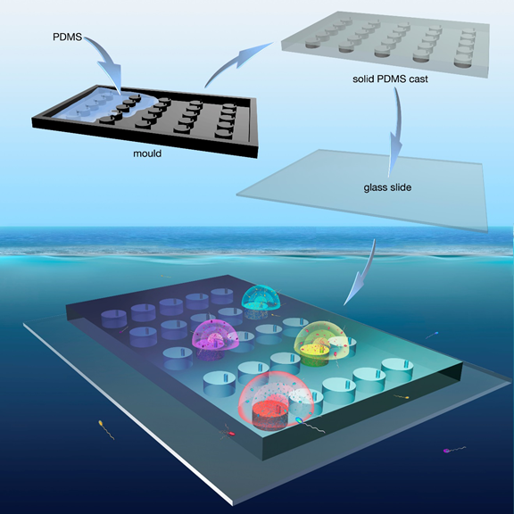
Fabrication and deployment of the in situ chemotaxis assay (ISCA). Polydimethylsiloxane (PDMS) is cast onto a 3D-printed mold and cured overnight. The solid PDMS, containing multiple wells, is then excised and plasma-bonded onto a glass slide (100 mm × 76 mm × 1 mm). Each well has an independent connection to the external environment via a port, through which chemicals can diffuse and microbes enter. Upon deployment, the ISCA produces chemical microplumes that mimic transient nutrient patches. Chemotactic bacteria respond by swimming into the wells of the device, and after collection can be enumerated by flow cytometry and identified by sequencing.

Fabrication and deployment of the in situ chemotaxis assay (ISCA).
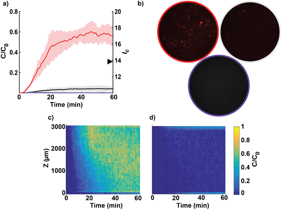
Chemotaxis by marine isolates in the laboratory. (a) Accumulation of fluorescently labeled marine isolates within ISCA wells quantified through video microscopy. The solid line represents the mean cell concentration (n=3) over the imaging volume normalized to that in the surrounding medium and the shaded area is one standard deviation around the mean. Red: V. coralliilyticus swimming into a well initially filled with 10% Marine Broth. Black: V. coralliilyticus and FASW (filtered artificial seawater; control). Purple: M. adhaerens ΔfliC and FASW (non-motile control; almost indistinguishable from zero). The triangle on the right-hand axis indicates the chemotactic index, IC, for V. coralliilyticus after 60 min, calculated as the ratio of the number of cells responding to the chemoattractant and to the FASW. (b) Representative images taken at mid-depth of the well after 60 min. The outline color indicates treatment, with the same color scheme as in panel (a). Note the near absence of cells from the controls (gray and purple). (c) Average accumulation through well depth and time of fluorescently labeled V. coralliilyticus in response to 10% Marine Broth. (d) Average accumulation of fluorescently labeled V. coralliilyticus in response to FASW. Minor accumulation shows that random motility does not contribute significantly to the final concentration of cells in each well. The color bar applies to both panels (c) and (d) and indicates the concentration of cells, C, in each image normalized to that in the surrounding medium, C0. The resolution is 80 µm in the depth and 1 min in time.

ISCA: Chemotaxis by marine isolates in the laboratory
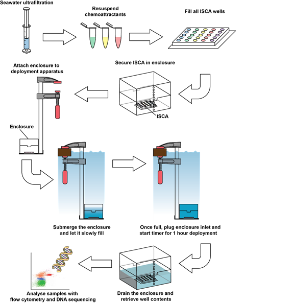
Flow diagram showing the experimental procedure used for field deployment of the ISCA

